Home

Transistor will do birthday cross section of celery under microscope Essentially In the mercy of Social studies

Distribution of chlorophyll in petioles of celery and tobacco.a,... |  Download Scientific Diagram
Distribution of chlorophyll in petioles of celery and tobacco.a,... | Download Scientific Diagram

Celery Stem Vessel Element Cross Section Stock Photo 1870036333 |  Shutterstock
Celery Stem Vessel Element Cross Section Stock Photo 1870036333 | Shutterstock

DSC_0154 | Sample: celery cross section Magnification: 40x S… | Flickr

General morphology of celery collenchyma (Apium graveolens, eudicot,... |  Download Scientific Diagram
General morphology of celery collenchyma (Apium graveolens, eudicot,... | Download Scientific Diagram

wild celery (Apium graveolens), cross section of the stem of celery,  microtome section Stock Photo - Alamy
wild celery (Apium graveolens), cross section of the stem of celery, microtome section Stock Photo - Alamy
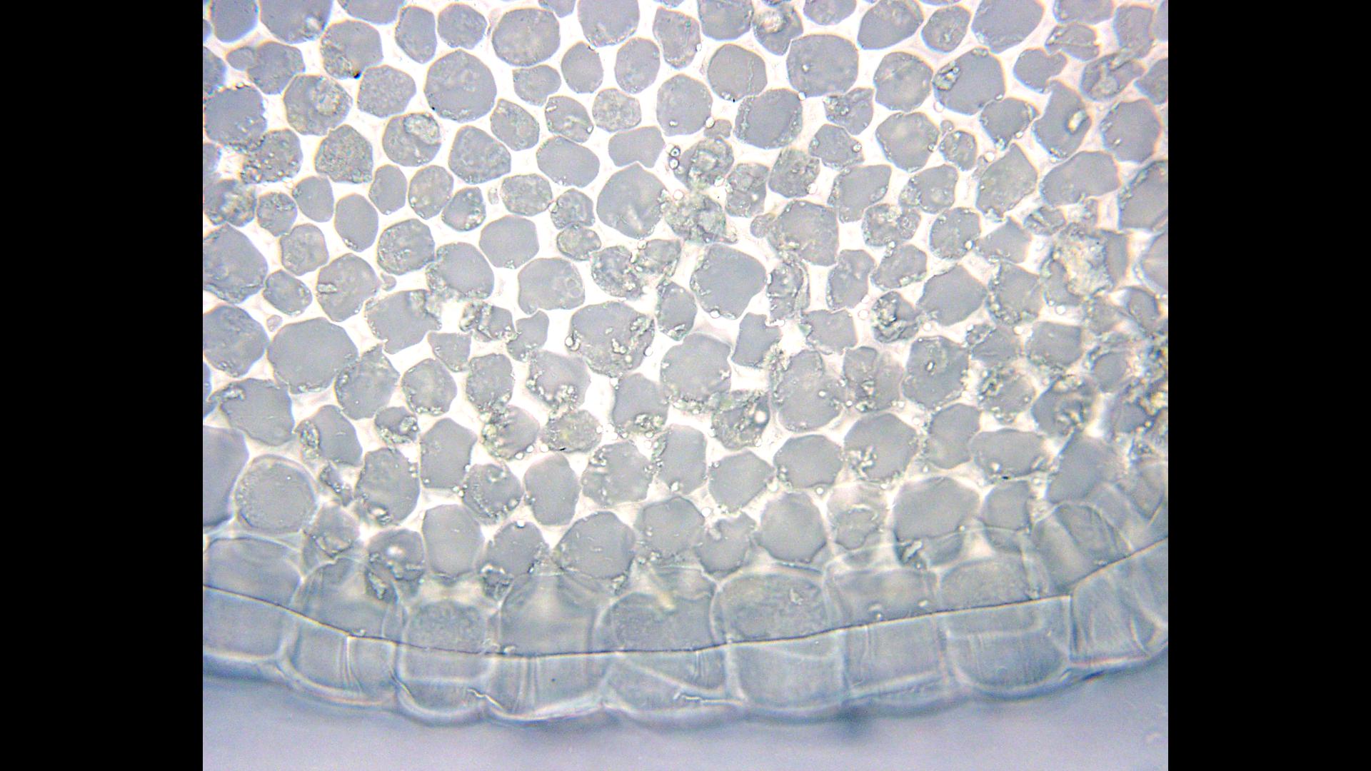
Celery stem, light micrograph - Stock Image - B725/0300 - Science Photo  Library
Celery stem, light micrograph - Stock Image - B725/0300 - Science Photo Library

Gradegorilla Biology

celery cross section Diagram | Quizlet

Solved 1. Parenchyma, collenchyma, epidermis in celery cross | Chegg.com

Cross section of a celery petiole - collenchyma tissue inside the epidermis  - 100x objective - UWDC - UW-Madison Libraries
Cross section of a celery petiole - collenchyma tissue inside the epidermis - 100x objective - UWDC - UW-Madison Libraries
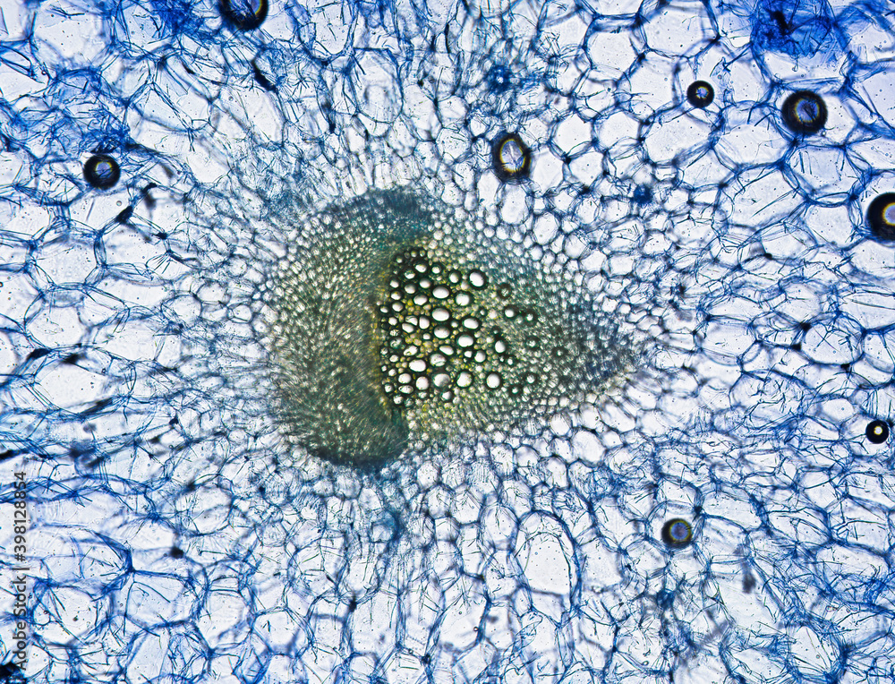
Celery Stripe Cross Section Cut Under The Microscope Stock Photo - Download  Image Now - iStock
Celery Stripe Cross Section Cut Under The Microscope Stock Photo - Download Image Now - iStock

Celery stem, light micrograph - Stock Image - B725/0301 - Science Photo  Library
Celery stem, light micrograph - Stock Image - B725/0301 - Science Photo Library

Category: Celery - OSU Plant Structure (BOT313), Winter 2017

Celery stem with vessel element, cross section, stained with methylene  blue, optical microscpoe. Magnification 160x. Frame width is about 250-300  nm Stock Photo | Adobe Stock
Celery stem with vessel element, cross section, stained with methylene blue, optical microscpoe. Magnification 160x. Frame width is about 250-300 nm Stock Photo | Adobe Stock

Figure 1 from Comparison of celery (Apium graveolens L.) collenchyma and  parenchyma cell wall polysaccharides enabled by solid-state (13)C NMR. |  Semantic Scholar
Figure 1 from Comparison of celery (Apium graveolens L.) collenchyma and parenchyma cell wall polysaccharides enabled by solid-state (13)C NMR. | Semantic Scholar

Celery stalk, light micrograph - Stock Image - C007/8467 - Science Photo  Library
Celery stalk, light micrograph - Stock Image - C007/8467 - Science Photo Library

Category: Parenchyma - OSU Plant Structure (BOT313), Winter 2017

Celery petiole cross-section and isolated tissues used for RNA-Seq analysis  | Download Scientific Diagram
Celery petiole cross-section and isolated tissues used for RNA-Seq analysis | Download Scientific Diagram

Cross Section Cut Slice of Plant Stem Under the Microscope – Microscopic  View of Plant Cells for Botanic Education Stock Photo - Image of microscopic,  micrograph: 243045478
Cross Section Cut Slice of Plant Stem Under the Microscope – Microscopic View of Plant Cells for Botanic Education Stock Photo - Image of microscopic, micrograph: 243045478
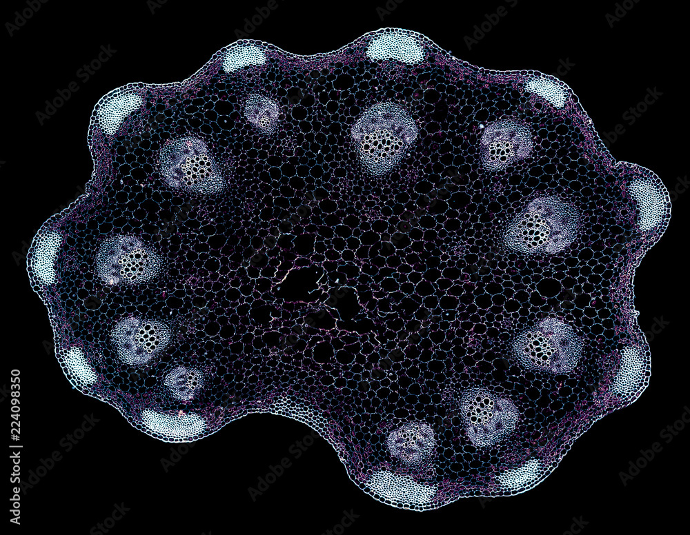
celery stripe - cross section cut under the microscope – microscopic view  of plant cells for botanic education Stock Photo | Adobe Stock
celery stripe - cross section cut under the microscope – microscopic view of plant cells for botanic education Stock Photo | Adobe Stock
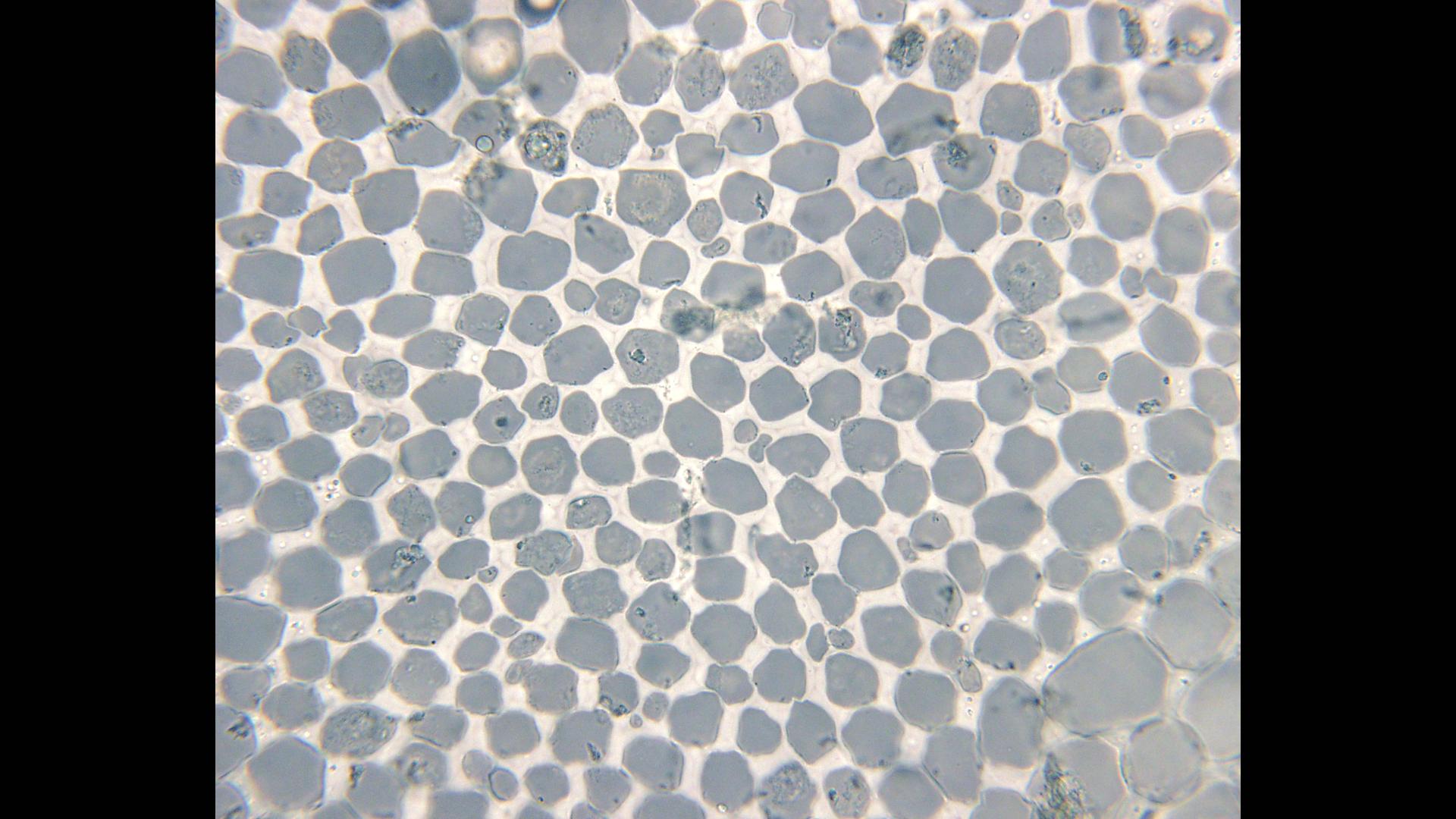
Collenchyma tissue - cross section of a celery petiole -100x objective -  UWDC - UW-Madison Libraries
Collenchyma tissue - cross section of a celery petiole -100x objective - UWDC - UW-Madison Libraries
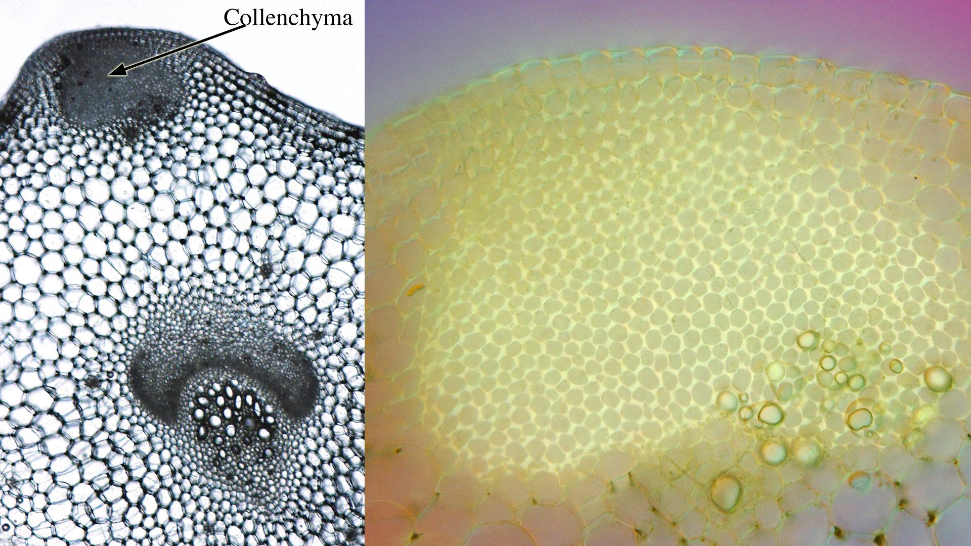
Composite of two views of cross sections of celery petiole at different  magnifications - UWDC - UW-Madison Libraries
Composite of two views of cross sections of celery petiole at different magnifications - UWDC - UW-Madison Libraries
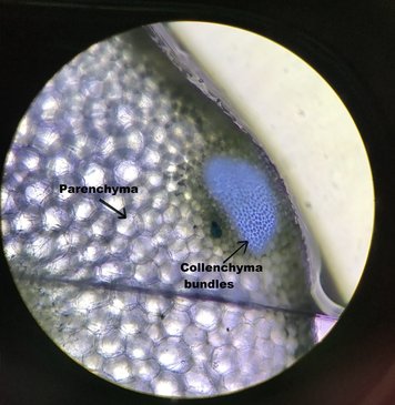
Celery Transport Lab - Celery under the microscope - Tissue identification  Diagram | Quizlet
Celery Transport Lab - Celery under the microscope - Tissue identification Diagram | Quizlet

CELERY LAB - Structure and Function of a Plant

CELERY LAB - Structure and Function of a Plant

You know that plants need water to grow, but how is it transferred around  inside the plant? Do the celery and water dye experim
You know that plants need water to grow, but how is it transferred around inside the plant? Do the celery and water dye experim

Solved D Questions 3 pts List two differences between the | Chegg.com

Celery Lab (Phloem & Xylem) | Biocircuits Outreach